Tuesday, 5 June 2012

What is Surgery?

According to Wikipedia, surgery is an ancient medical specialty that uses operative manual and instrumental techniques on a patient to investigate and/or treat a pathological condition such as disease or injury, or to help improve bodily function or appearance. There are many specific fields/specialties of surgery and surgeons. Some of the most famous fields include cardiac/cardiothoracic surgery, which deals with the heart and lungs, neurosurgery, which deals with the brain and the nervous system, orthopedic surgery, which deals with bones and joints, ENT surgery, which deals with the ears, nose, and throat, and plastic surgery, which deals with the appearance of a human being. Here is a diagram that shows a visualization of a open heart surgery, where the chest is opened and the sternum is cut.
external image open-heart-surgery-incision-diagram.jpg
Here is a diagram of a craniotomy, which is a form of brain surgery, where the surgeon removes part of the skull bone and cuts into the dura mater.
external image craniotomy_2.jpg

Surgical Instruments

Surgeons use many tools, called surgical instruments, to operate on a patient, in an operating room (O.R.). A typical OR at a hospital looks like this.
 external image ms_pic_ohiostatenew-lowrez1.jpg
It includes bright lights, a table that can be tilted any way, monitors that show a camera above the patient, and an instrument tray, with various doctors and nurses working. A scalpel is used to cut, into the skin, blood vessels, etc.
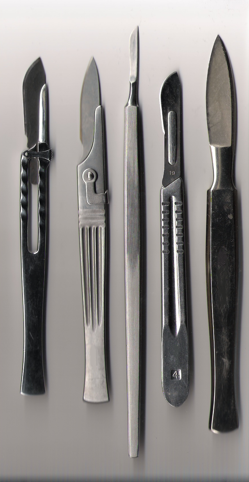
 external image Various_scalpels.png

Scissors are used to cut, like scissors are meant to be used. They can be used to cut through muscle, wires, umbilical cords, etc.
 external image 1316.jpg
Clamps are used to apply pressure and cut off circulation. This is an aortic cross-clamp. external image paediatric.jpg

Rib spreaders are used to separate the ribs at the sternum after cutting it, for an open heart surgery. external image l_7033.jpg
Retractors are used to widen the incision so the surgeon can further manipulate or see into the internal organs. These are Roux retractors.
external image 22-1600.png

Those are only some of the equipment and instruments used by surgeons. There are much more that aren't listed here.

Open Heart Surgery

Open cardiac/heart surgery is a type of surgery in which the surgeon makes an incision on the skin above the sternum, then uses a bone saw to cut through the sternum in order to have direct access to the heart, for transplantation, or other procedures. In major procedures, the surgeon may attach the patient to a heart-lung machine, also called a bypass, and stop the heart in order to work on it, such as in valve replacements.
Here is video on an aortic valve replacement.


The bone saw looks like this.

external image Surgical-Sternum-Saw-RJ02006-.jpgexternal image 78-A.jpg

Heart-Lung Bypass

Bypass is used when the surgeon needs to stop the heart to work on it. Tubes are attached from the bypass machine into the major arteries. The patient is intubated to help him/her breathe. The machine will pump the blood and breathe for the patient. This way, the heart can be stopped and blood will continue circulating the body. A heart lung bypass machine looks like this.

external image BypassMachine.jpg

As you can see, there are many complicated tubes coming out it that help circulate blood. It is also connected to a packet of blood of the same type as the patient.

Craniotomy

A craniotomy is when a neurosurgeon drills into the skull of a patient in order to get access to their brain to remove a tumor, relieve pressure, or other reasons.

The procedure starts with taking a CT or MRI scan of the patient's head. This helps the surgeon precisely locate the angle where he has to cut and the location. Then, the head is prepared. The neurosurgeon will cut the hair around the area where the surgery will take place. Then, the surgeon will cut into the scalp and hit the skull. The high tech neurosurgery drill will be used to remove part of the skull flap. It will be replaced later.

This is a high tech neurosurgery drill.
external image images?q=tbn:ANd9GcQm2heVT4FmR3iosP0SGFcq2HJKcPFl_MnaAG9-Mm1gNNAbFKdK240ib7fP
Here are some instruments designed for precision in neurosurgery.
external image Instruments_large.jpg


Finally, here is a video of a craniotomy on the right parietal lobe of the brain.

Appendectomy

An appendectomy is the removal of the vermiform appendix. The appendix is located at the tip of your large intestine. This procedure can be done laporascopically, or as an open surgery.
The procedure for an open appendectomy is as follows:

  1. Antibiotics are given immediately if there are signs of sepsis, otherwise a single dose of prophylactic intravenous antibiotics is given immediately prior to surgery.
  2. General anaesthesia is induced, with endotracheal intubation and full muscle relaxation, and the patient is positioned supine.
  3. The abdomen is prepared and draped and is examined under anesthesia.
  4. If a mass is present, the incision is made over the mass; otherwise, the incision is made over McBurney's point, one third of the way from the anterior superior iliac spine (ASIS) and the umbilicus; this represents the position of the base of the appendix (the position of the tip is variable).
  5. The various layers of the abdominal wall are then opened.
  6. The effort is always to preserve the integrity of abdominal wall. Therefore, the External Oblique Aponeurosis is slitted along its fiber, and the internal oblique muscle is split along its length, not cut. As the two run at right angles to each other, this prevents later Incisional hernia.
  7. On entering the peritoneum, the appendix is identified, mobilized and then ligated and divided at its base.
  8. Some surgeons choose to bury the stump of the appendix by inverting it so it points into the caecum.
  9. Each layer of the abdominal wall is then closed in turn.
  10. The skin may be closed with staples or stitches.
  11. The wound is dressed.
  12. The patient will be brought to the recovery room.